Play: Spot the Finding

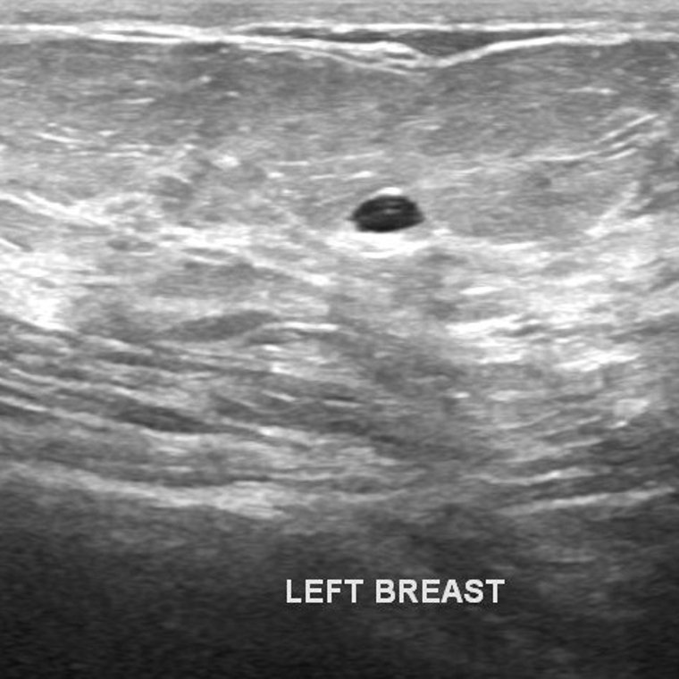
A quick, honest way to feel what these specialists do. You are shown a real breast image from one of the modalities; decide whether it looks benign / normal or suspicious — refer. The AEGIS engine then reveals what to look for. It is harder than it sounds — which is exactly why a screening aid, and a human in command, help.

Ultrasound
Breast screening image to classify Real de-identified research image · BUSI / BACH / CBIS-DDSM
Benign or suspicious?
Score: 0 / 0

Game images are real, de-identified samples from public research datasets — breast ultrasound (BUSI), histology microscopy (ICIAR-2018 BACH), and mammography (CBIS-DDSM) — shown for education only, never a diagnosis. Ground-truth labels are the datasets’ own. The decorative banner at the top is an AI-generated illustration.

What this is

The AEGIS Unified Breast Station brings every AEGIS breast specialist under one roof and routes each modality to the specialist that reads it. It runs entirely on a local machine — no image leaves it — and it is honest by construction: each specialist stands on its own validated metric, and any case that lands in the grey zone is flagged for a clinician, never auto-called. AEGIS scores; a clinician confirms.

Read this first — what is built, and what it is not. This is a feasibility research programme: a bench of single-modality screeners, each trained and validated on a public research dataset, unified by one routing engine. Every number below is an honestly-reported validation AUC on held-out data — not a clinical-trial result. AEGIS Breast does not diagnose. It is decision support with a human always in command.

The specialists — honest metrics

Each row is a separate validated specialist. The strong diagnostic-grade readers clear the conventional 0.90 floor; the weaker ones are reported as weaker, with the reason, rather than dressed up — that honesty is the point.

SpecialistModality / inputChampionValidation ROC-AUC
DIAGWDBC fine-needle-aspirate (30 nucleus measures)Fuzzy Linear-SVC0.9955
USBreast ultrasound (BUSI)Nu-SVC0.9328
BreaKHisTissue microscopy (patient-separated)Fuzzy SVM-RBF0.9302
BACHHistology microscopy (4-class carcinoma)Nu-SVC0.9048
HISTHistology H&E patch (PatchCamelyon)Logistic Regression0.9014
MAMMMammogram (CBIS-DDSM, whole-film @224px)Logistic Regression0.7149 (screening baseline — see note)
PROGWPBC prognostic measures (recurrence, n=198)Logistic Regression0.6595 (risk aid, not a gate)

Honest notes: MAMM 0.71 is a legitimate whole-film screening baseline — malignancy is a small region of a large mammogram, and downscaling to 224px sheds the fine calcification detail; ROI-cropped patches at higher resolution are the lever to lift it. PROG 0.66 is recurrence prediction on 198 single-source patients — genuinely hard, so it is a risk aid with a caveat, never a diagnostic gate. HIST, DIAG, US, BACH and BreaKHis are the real diagnostic-grade gates.

We’re not breast specialists — help us make AEGIS better through more in-depth training

Honestly: AEGIS is a research programme, not a clinic, and we’re not radiologists or pathologists. The system improves as it sees more real examples. You can already try it right now — the “Spot the Finding” station above runs on real de-identified images from public research datasets, so you can test AEGIS without sending anything. And if you’d be willing to share a de-identified breast image, you’d be helping us test and sharpen it — and we’ll show you what the model sees, as an illustration only, never a diagnosis. A few honest ground rules first; sending an image means you accept them. Thank you for your support!

A few honest ground rules before you send anything.
1. Not a medical device and not a diagnosis. AEGIS is a research and educational demonstration. Nothing it outputs is medical advice, a diagnosis, a screening result, or a substitute for a qualified clinician. No doctor–patient relationship is created.
2. Do not send identifiable patient data. Remove all names, IDs, dates of birth, and any protected health information (PHI) before sending. Only submit an image you have the right to share. Do not send anyone else’s medical data without their consent.
3. You release AEGIS from liability. By submitting, you agree to indemnify and hold harmless AEGIS, Dr Loh Kah Meng, and anyone associated with the programme from any and all claims, losses, or liability arising from your use of, or reliance on, anything AEGIS provides. You use it entirely at your own risk.
4. Always consult a licensed clinician. For any real health concern, see a qualified doctor. Do not delay or disregard professional medical advice because of anything here.

This opens your email app to [email protected] with the terms pre-acknowledged. Attach your de-identified image before sending.

One programme, one honest promise. AEGIS augments, it never replaces. Every specialist reports its own real number; grey-zone cases defer to a person; nothing is auto-called; and no work is dressed up beyond what the data supports. Dedicated to Professor Dhanjoo N. Ghista, whose nondimensional-index lineage this work continues.
📬 Get new-app alerts Contact us