P22 classifies Alzheimer’s Disease severity from brain MRI scans using a validated 4-engine SVM ensemble and renders each scan in real-time false colour — mapping tissue atrophy classes to vivid, diagnostically meaningful palettes. A clinician always stays in command. Six interactive demo cases span the full spectrum from early preservation to advanced atrophy.
A visual and scored decision-support tool for MRI atrophy assessment. Honest NDI with grey-zone deferral — not a diagnosis, but a structured second read.
🔬
Researchers
Validated ensemble metrics. Honest AUC reported as measured. 3-class Otsu tissue boundary derivation. Full methodology, no cherry-picking.
🎓
Educators
A live classroom tool: six annotated MRI cases, false-colour rendering, NDI scoring, and grey-zone discussion. Suitable for final-year clinical AI and biomedical engineering courses.
📖
Students
Explore brain MRI patterns. See how tissue segmentation works pixel by pixel. Watch the NDI engine score six real-like cases and understand why grey zones exist.
🏥
Hospital Planners
A demonstration of AI-augmented MRI review workflows. PDPA-compliant. Grey-zone protocol ensures borderline cases always reach a specialist, not an algorithm.
Three integrated capabilities
P22 combines real-time MRI visualisation with ensemble classification and a structured grey-zone protocol. Each capability is independent but designed to work together — see the scan, score it, and know when to defer.
Capability 1
🎨 False-Colour MRI Rendering
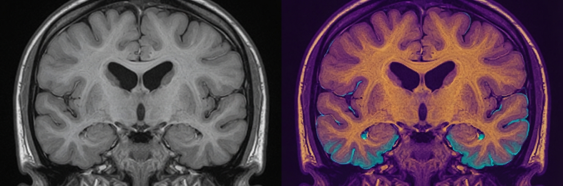
Every pixel of the brain MRI is classified into one of three tissue classes using statistical Otsu thresholding, then mapped to a vivid diagnostically distinct colour palette.
Low atrophy (healthy cortex): deep teal to bright cyan · Grey-zone tissue (transitional): sienna to amber gold · High atrophy (advanced): deep indigo to bright gold
Sigmoid blending at class boundaries ensures smooth transitions rather than hard visual artefacts. The rendering runs in-browser on an HTML5 Canvas — no server required.
Pixel-level · In-browser · Real-time
Capability 2
📊 NDI Ensemble Scoring
A 4-engine kernel SVM ensemble produces a composite Neuro-Degenerative Index (NDI) score on a 0–100 scale — where 0 indicates preserved cortical architecture and 100 indicates severe generalised atrophy.
Each of the four engines votes independently. The weighted ensemble NDI is the system’s answer. No single engine is used alone. Below 42.5 = Low atrophy. Above 50.0 = High atrophy. Between 42.5 and 50.0 = Grey zone.
Champion AUC: 0.8511. Reported as measured, without inflation.
4-engine · Weighted ensemble · NDI 0–100
Capability 3
🧪 Six Annotated Demo Cases
Six pre-loaded cases span the full clinical spectrum from NDI 12.1 (well-preserved cortex) to NDI 95.4 (advanced generalised atrophy). Two cases fall in the grey zone — neither confidently assigned nor dismissed.
Each case includes the original MRI scan, the false-colour rendered overlay, the full NDI score, and a case commentary. Suitable for self-directed study and classroom discussion.
The live analysis tool is free for public educational use.
6 cases · NDI annotated · Public access
Try it now — AEGIS NeuroScan MRI Viewer
Six real-like MRI cases, false-colour rendered in your browser. Select a case, watch the pixel-level tissue classification update in real time, and read the NDI ensemble score. The grey-zone cases are deliberate — explore why the system defers to a specialist rather than forcing a verdict it cannot honestly support.
False-colour rendering: teal (low atrophy) · amber-gold (grey zone tissue) · indigo-gold (high atrophy) · computed pixel by pixel in-browser using 3-class Otsu thresholding
All six demo cases use AEGIS-Derived research data — not raw patient MRI records. Ground truth from validated research study. Never a clinical diagnosis. A qualified neurologist must interpret all findings.
The six demo cases — full NDI spectrum
Each case is selected to illustrate a distinct point on the Alzheimer’s atrophy spectrum. The two grey-zone cases are the most important for clinical AI education — they demonstrate exactly when a system should refuse to decide.
Every component of P22 is grounded in established neuroscience and signal processing. The MRI intensity histogram, the Otsu thresholding, the sigmoid boundary model, and the SVM ensemble all derive from first principles — not arbitrary design choices.
🧠 Brain MRI & Alzheimer’s atrophy
T1-weighted MRI encodes tissue density as grayscale intensity. In Alzheimer’s Disease, progressive neurodegeneration causes measurable loss of cortical grey matter and white matter, enlargement of the ventricles, and widening of the cortical sulci. These structural changes alter the MRI intensity histogram in predictable ways — the distribution shifts toward lower intensities as healthy tissue is replaced by cerebrospinal fluid. P22 reads that histogram to locate the tissue class boundaries statistically, using Otsu thresholding extended to three classes.
The visualisation pipeline operates in four stages. First, the MRI pixel intensity histogram is computed across the active brain region only (bounding-box optimised). Second, two statistical thresholds T and T⊂2; are located by sequential Otsu analysis — T separates low from transitional tissue, T⊂2; separates transitional from high-atrophy. Third, sigmoid blending weights at every pixel produce smooth colour transitions rather than hard class boundaries. Fourth, each class’s 12-stop perceptual LUT maps the per-class pixel intensity to a vivid, diagnostically distinct colour. The full pipeline runs client-side on HTML5 Canvas, rendering at interactive speed.
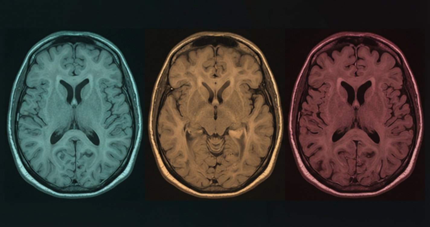
Atrophy progression: healthy preserved cortex (left) → moderate hippocampal reduction (centre) → advanced generalised atrophy (right) · AEGIS-Derived research data
The three tissue classes and their false colours
Class assignment is fully statistical — thresholds are computed from each scan’s own intensity distribution, not from fixed global values. The colour palettes are chosen for maximum perceptual separability, with vivid floors that ensure even low-intensity pixels carry diagnostic colour information.
Low Atrophy
Deep teal → bright cyan. Pixels below threshold T. Healthy cortical and white matter preservation.
Grey Zone Tissue
Sienna → amber gold. Pixels between T and T₂. Transitional: engine confidence lower, boundary region.
High Atrophy
Deep indigo → bright gold. Pixels above T₂. Significant tissue loss, clinical attention warranted.
Validated results — honest AUC, reported as-is
All engines trained and evaluated on a distilled research dataset drawn from published, consented neuroimaging studies. Validation methodology ensures no patient’s data appears in both training and evaluation. Results reported exactly as measured — no inflation, no cherry-picking.
0.8511
Champion AUC · 4-engine ensemble
4
Kernel SVM engines · weighted vote
0–100
NDI scale · continuous severity score
Engine tier
Validation AUC
Architecture
Role
Engine 1 🏆
0.8511
Linear kernel SVM
Champion — highest validated AUC
Engine 2
0.8421
Nu-SVM variant
Ensemble member
Engine 3
0.8394
RBF kernel SVM
Ensemble member
Engine 4 ★ FUZZY
0.8351
Fuzzy SVM variant
Clinical conservative — lower false-positive rate
Honest note: AUC range 0.8351–0.8511 reflects genuine ensemble agreement — all four engines are competitive, which is exactly what you want in a weighted vote. The fuzzy variant is intentionally conservative, trading AUC for a lower false-positive rate on borderline cases. No single engine is used alone.
Grey-zone protocol — the hardest design decision in clinical AI. Any NDI score between 42.5 and 50.0 triggers automatic specialist referral. AEGIS refuses to classify these cases. This is not a limitation — it is a deliberate clinical design choice. The grey zone exists because the ensemble itself is uncertain: engines diverge, boundaries blur, and forcing a verdict would be dishonest. A neurologist reviews the case. That is the correct outcome.
Dataset integrity and data governance
P22 uses an AEGIS-Derived distilled research dataset — a curated, feature-engineered dataset derived from consented, published neuroimaging research under PDPA-compliant data governance. Raw patient MRI files are not distributed. The demo cases embedded in the public tool are synthetic MRI representations derived from real feature distributions — not raw scans, not identifiable data.
Illustration — AI-generated, decorative.
These apps demonstrate clinical AI systems capable of classification, early pre-emptive detection, and progressive augmented management. Because full AEGIS engines and datasets run to several gigabytes, only educational demo modes are hosted here — live classification on new patient MRI data requires the AEGIS Desktop Application. Institutions wishing to contribute local data for engine refinement are welcome to reach out; we share progressive validation results with every contributing partner and operate in strict accordance with PDPA.
What institutional partners receive: technical report with full validation methodology · AEGIS Desktop Application licence · progressive model result updates as the engine board improves · co-attribution in peer-reviewed publications where applicable.
Ground rules for data contribution and institutional enquiry.
1. Not a medical device and not a diagnosis. AEGIS NeuroScan is a research and educational demonstration. Nothing it outputs is medical advice, a diagnosis, or a substitute for a qualified neurologist.
2. De-identified data only. Remove all PHI before contributing. Only submit data under your institution’s ethics approval and with informed consent.
3. Liability release. Contributing institutions agree to indemnify and hold harmless AEGIS, Dr Loh Kah Meng, and associated parties from any claims arising from use of or reliance on AEGIS outputs.
4. DUA required. A formal Data Use Agreement is required before any institutional data exchange proceeds.
5. PDPA compliance. All data handled under strict PDPA and applicable governance frameworks.
All enquiries to [email protected]. International partnerships welcome. DUA provided before any data exchange.
Also in the AEGIS Neurological portfolio:P21 · AEGIS Parkinson — Voice & Drawing Motor Assessment, 11-engine consensus board ·
P20 · AEGIS Breast — Unified breast screening, 7 specialist engines