Who this is for
Neurologists & Radiologists
A confidence-zoned decision-support reference for brain MRI review. Four-zone routing surfaces cases requiring specialist deferral before any clinical action is taken.
Researchers
Validated ensemble metrics on a distilled MRI research dataset. Honest per-class accuracy reported as-is. 15-engine AEGIS ensemble with binary and 4-class outputs.
Educators
A live classroom tool: 16 annotated MRI cases across all four tumour classes and all four confidence zones — demonstrating AI-augmented radiology for BME students and clinical AI courses.
Students
See how MRI features translate to AI classification. Explore how a multi-engine ensemble distributes probability across Glioma, Meningioma, Pituitary, and No Tumour categories.
Hospital Planners
A demonstration of AI-augmented brain tumour triage. Zero PHI storage. Zone protocol ensures borderline cases receive mandatory human review before any workflow action.
Clinical AI Developers
A reference implementation of 4-zone confidence routing applied to MRI classification — a design pattern for responsible AI deployment in high-stakes medical imaging contexts.
Three integrated capabilities
P24 combines confidence-zone governance with a four-class MRI tumour classifier and a binary tumour-detection screen, all running through the 15-engine AEGIS AI ensemble on every case.
🚦 4-Zone Confidence Triage
Every MRI case is routed to one of four governance zones based on the ensemble's classification confidence. Zone assignment drives the clinical recommendation — from VERIFIED (act) through LOW (mandatory specialist deferral).
🧬 4-Class MRI Classification
The AEGIS AI ensemble classifies across all four brain tumour categories — Glioma, Meningioma, Pituitary Tumour, and No Tumour — with 99.03% validated accuracy on the distilled research dataset.
⚡ Binary Tumour Detection
An independent binary screen separates Tumour from No Tumour across all 7,200+ training MRI images, validated at AUC 0.9997 — the highest-confidence output in the system for preliminary screening.
Try it now — AEGIS TumorSentinel Demo Console
16 pre-loaded MRI cases spanning all four tumour classes and all four governance zones. Each case shows the full ensemble output: class, confidence percentage, binary screen result, zone assignment, and the clinical recommendation the zone drives.
16 pre-loaded demo cases — 4 tumour classes × 4 confidence zones. Click any case to see the full AEGIS AI ensemble result instantly. Upload your own MRI in Live Mode to generate a shareable retrieval code.
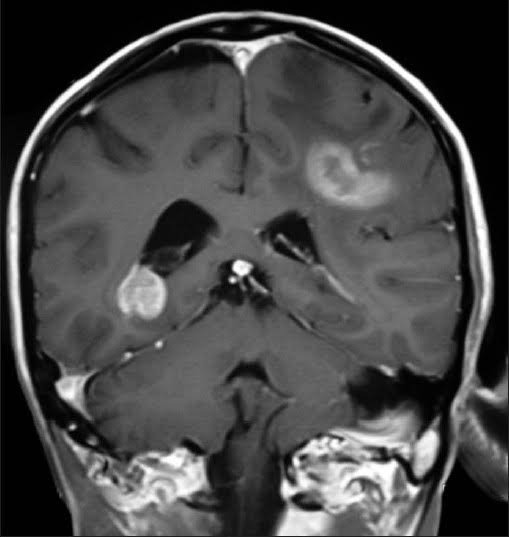
VERIFIED
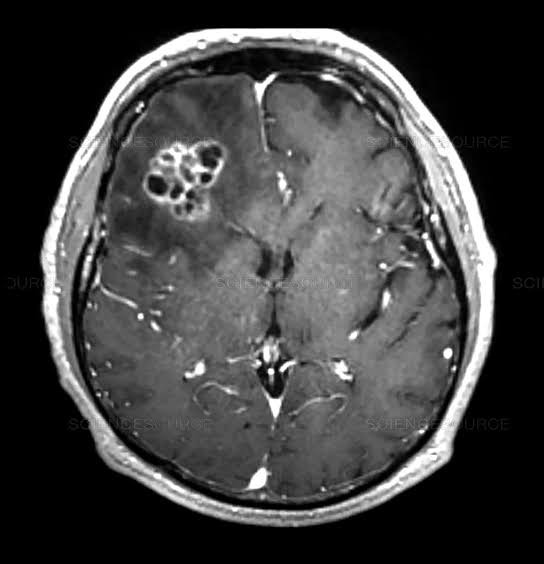
HIGH
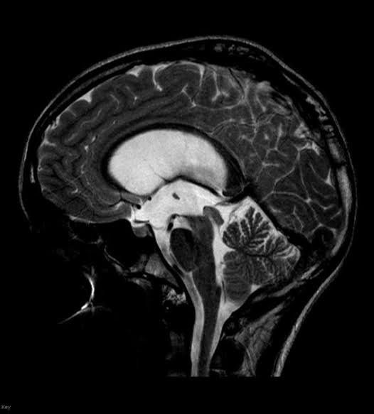
MID
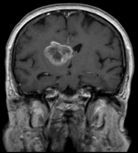
LOW
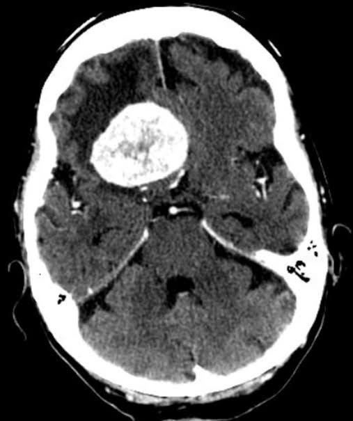
VERIFIED
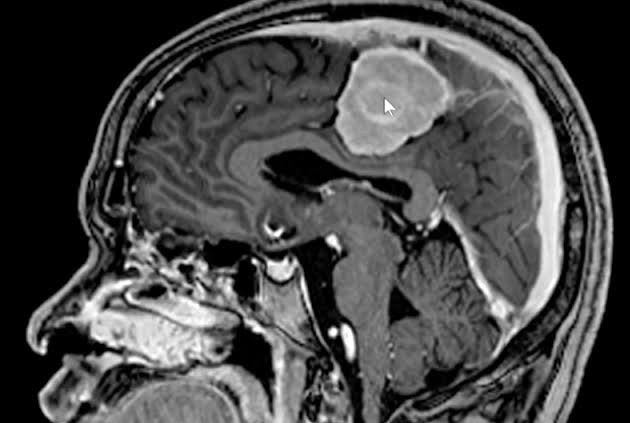
HIGH
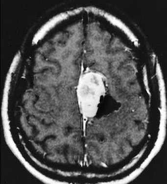
MID
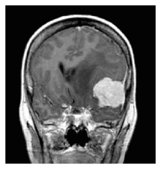
LOW
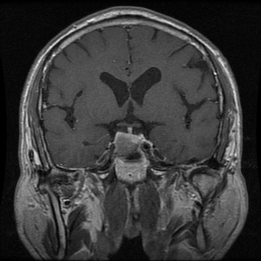
VERIFIED
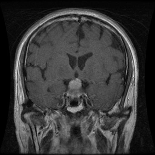
HIGH
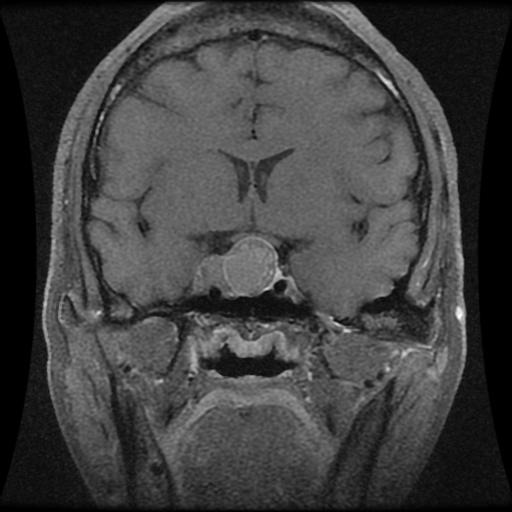
MID
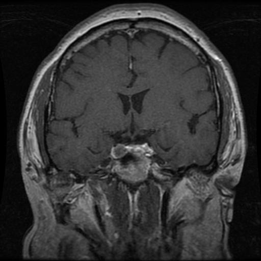
LOW
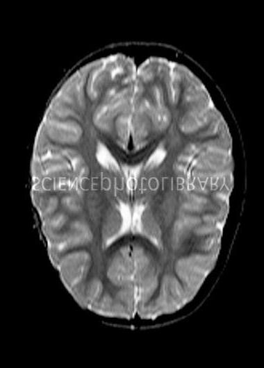
VERIFIED
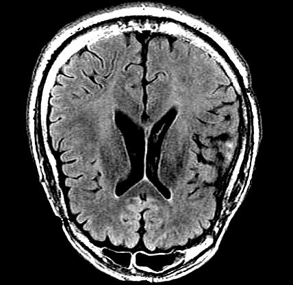
HIGH
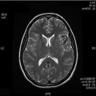
MID
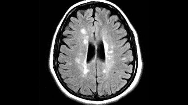
LOW
All demo cases use synthetic pre-set values drawn from the research dataset. No real patient data. Zero PHI storage.
The four governance zones
Zone assignment is computed from the AEGIS ensemble's classification confidence. The four zones map ensemble confidence to a specific clinical recommendation — protecting patients by ensuring low-confidence cases are never acted on without specialist review.
High Confidence
Ensemble strongly supports the primary classification. Result can be used to inform the clinical workflow. Clinician confirms before action.
Good Confidence
Ensemble reasonably supports the classification but with lower certainty. Radiologist notification recommended before acting on the result.
Elevated Uncertainty
Ensemble uncertainty is elevated. Human review required before any clinical decision. Do not use the classification as a primary determinant.
Defer — Specialist Required
Ensemble confidence is insufficient for any clinical use. Mandatory immediate specialist referral. The result must not drive any clinical decision.
The four brain tumour classes
AEGIS P24 classifies across all four categories from the brain tumour MRI research dataset, spanning malignant primary tumours, benign tumours, and normal (no tumour) scans.
Glioma
Primary brain tumour arising from glial cells. Includes GBM (Grade IV), the most aggressive form. Irregular enhancement pattern on MRI. Any Glioma result requires immediate neurosurgical review.
Meningioma
Arises from the meninges surrounding the brain and spinal cord. Usually slow-growing; majority benign (WHO Grade I) but higher grades exist. Often presents with dural tail sign on MRI.
Pituitary Tumour
Adenoma of the pituitary gland. Typically benign with characteristic sellar/suprasellar location on MRI. Endocrine consequences may require management independent of malignancy classification.
No Tumour
No tumour mass identified in the MRI scan. Validated against 7,200+ MRI images including normal scans. Binary AUC 0.9997 for Tumour vs No Tumour detection.
The biomedical engineering science
P24 is grounded in neuroimaging science, deep learning transfer, and clinical AI safety design. The system uses a 15-engine AEGIS AI ensemble trained on distilled datasets derived from publicly available brain MRI research data.
🧠 Brain MRI and tumour classification
Brain MRI distinguishes tumour subtypes through signal intensity, contrast enhancement patterns, mass effect, and anatomical location. T1 post-contrast, T2, and FLAIR sequences carry distinct diagnostic weight. The AEGIS P24 training corpus spans axial MRI slices across all four categories.
⚙️ AEGIS AI ensemble pipeline
P24 runs a 15-engine AEGIS AI ensemble drawing on deep learning feature extraction and ensemble inference across multiple algorithmic approaches. Engine identity, weights, and architecture are IP-protected under the AEGIS research framework. The output is a class probability distribution and confidence score.
📐 Adaptive governance zone routing
Confidence scores are routed through a zone classifier that assigns each result to VERIFIED, HIGH, MID, or LOW — with each zone carrying a specific clinical recommendation. Zone thresholds are class constants defined in the research framework.
🔒 Zero PHI storage
The web application performs all inference client-side. No MRI image or patient identifier is transmitted or stored. Retrieval codes are self-decoding tokens containing only the classification result — no imaging data.
Validated results — honest accuracy, reported as-is
All engines trained and evaluated on distilled datasets drawn from the brain tumour MRI research corpus under strict train/test separation. Results reported as measured.
| Task | System | Metric | Value | Note |
|---|---|---|---|---|
| Binary detection | AEGIS AI ensemble (15 engines) | AUC | 0.9997 | Tumour vs No Tumour · distilled research dataset |
| 4-class classification | AEGIS AI ensemble (15 engines) | Accuracy | 99.03% | Glioma / Meningioma / Pituitary / No Tumour |
| Training corpus | — | MRI images | 7,200+ | Brain tumour MRI research dataset |
Dataset integrity and data governance
P24 is trained and validated on a distilled research dataset derived from the brain tumour MRI public research corpus — a widely used benchmark in neuro-oncology AI research.
Not a medical device
AEGIS TumorSentinel is a research and educational demonstration. Nothing it outputs is medical advice. Results must not substitute for clinical assessment by a qualified neurologist or radiologist.
De-identified data only
No real patient data was used in training or demo. All demo cases are pre-set synthetic values. No MRI is stored on any server. The system processes images locally in the browser.
PDPA compliance
Zero PHI storage architecture. Retrieval codes contain only classification meta — no imaging data. Institutional use requires a Data Use Agreement before any data exchange.
Institutional partners receive
Full technical report with validation methodology · AEGIS Desktop Application licence · Progressive clinical trial protocol · DUA provided before any data exchange.
All enquiries to [email protected]. International partnerships welcome. DUA provided before any data exchange.